3 Questions to Ask About X-Ray Services
X-rays are useful for diagnosing bone and soft tissue irregularities such as fractures, dislocations, and osteoporosis due to density variations. They are useful for diagnosing diseases, determining treatment options, tracking progression, and evaluating treatment effectiveness. At PACS, Dr. Khaled Said MD, and Dr. Walid Hammad, offer X-ray services. For more information, contact us today or schedule an appointment online. We have convenient locations to serve you in Alexandria VA, and Ruther glen VA.


Table of Contents:
What is X-ray imaging used for?
Is X-ray the same as imaging?
What is the difference between an X-ray and a radiograph?
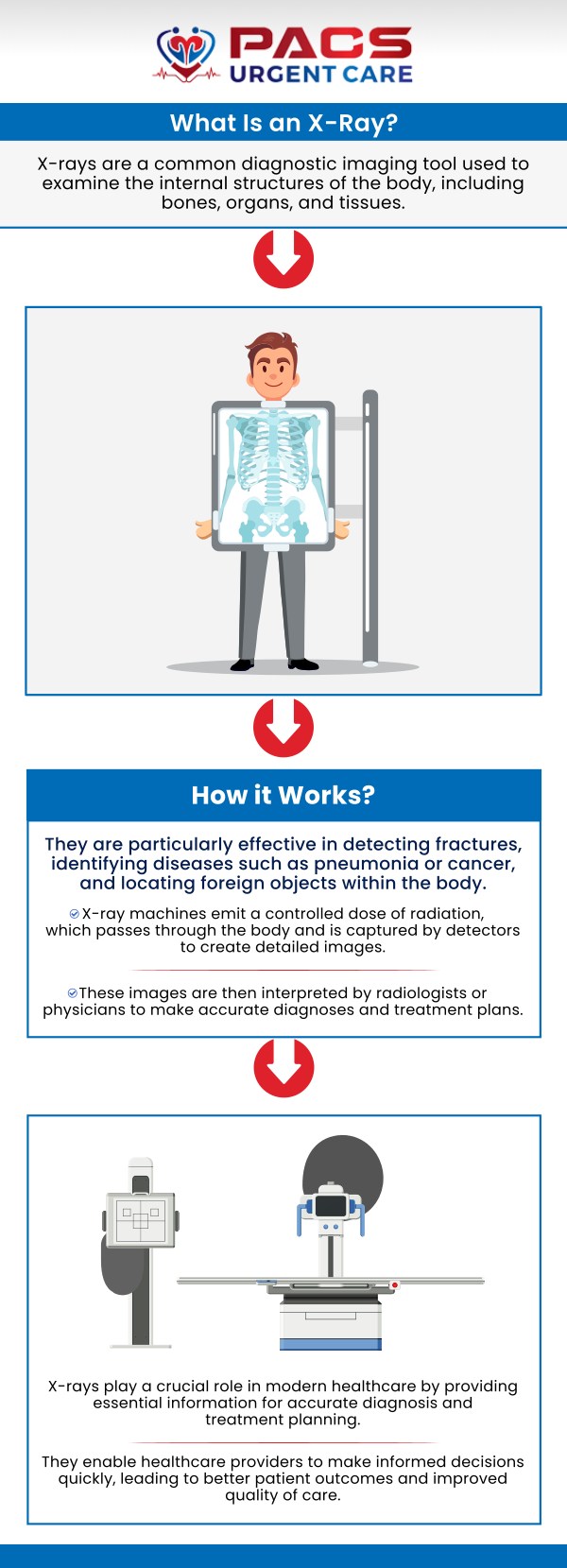
X-ray imaging, also known as radiography, is a diagnostic tool mostly seen and used in medical offices to see the inner structures of the body. It works by passing a small amount of ionizing radiation through the body to create images on a film or digital sensor. X-rays are particularly effective in detecting abnormalities in bones, such as fractures, dislocations, or osteoporosis, due to differences in their density.
They can also identify certain soft tissue abnormalities, such as pneumonia in the lungs, kidney stones, or tumors. X-ray images can provide valuable information for diagnosing a range of conditions, guiding treatment decisions, monitoring progression, and assessing the effectiveness of treatments. While X-rays are an essential and widely used imaging modality, they have limitations in visualizing soft tissues, such as muscles and organs, and cannot diagnose all conditions. Other imaging techniques, like computed tomography (CT) scans, magnetic resonance imaging (MRI), or ultrasound, may be utilized in conjunction with X-rays to provide a more comprehensive evaluation of the body. It is imperative to think about the potential risks associated with ionizing radiation exposure when ordering X-rays, especially for pregnant individuals or children. Despite these considerations, X-ray imaging remains a valuable tool in healthcare for diagnosing and monitoring a variety of medical conditions with precision and accuracy.
X-ray imaging is a subset of medical imaging, a field that encompasses various techniques for visualizing anatomical structures within the body. While X-ray imaging uses ionizing radiation to capture images of bones, tissues, and organs, it is not the only imaging modality available in healthcare. Computed tomography (CT) scanning uses X-rays to create cross-sectional images of the body, providing detailed information on organs, soft tissues, and bones. Magnetic resonance imaging (MRI) can use magnetic and radio frequencies to generate detailed images of structures like the brain, spine, and joints, offering valuable insights into neurological, musculoskeletal, and soft tissue conditions without the use of ionizing radiation.
Ultrasound imaging typically consists of high-frequency sound waves that make real-time images of the organs and tissues within the body, making it particularly useful for evaluating the heart, blood vessels, and developing fetus during pregnancy. Nuclear medicine imaging, on the other hand, involves the injection of radioactive substances into the body to detect abnormalities in organ function, metabolism, and blood flow. Each imaging modality has its unique strengths, limitations, and applications in diagnosing and monitoring a wide range of medical conditions. Collectively, these modalities contribute to the comprehensive field of medical imaging, playing a vital role in modern healthcare by assisting in the diagnosis, treatment planning, and monitoring of various diseases and injuries.
An X-ray and a radiograph are fundamental components of medical imaging that play a crucial role in diagnosing and monitoring a wide range of medical conditions. X-rays are used to visualize internal structures within the body by passing X-ray beams through the body and capturing the remaining rays on a detector. The resulting image, known as a radiograph, displays the absorption pattern of X-rays by different tissues, such as bones, organs, and soft tissues. In a radiograph, bones appear as white structures due to their higher density and ability to absorb more X-rays, while soft tissues appear as shades of gray, and air-filled spaces appear as black areas. The detailed information obtained from radiographs helps healthcare professionals identify fractures, tumors, infections, and other abnormalities that may not be visible externally.
Radiographs are commonly used in various medical specialties, including orthopedics, dentistry, emergency medicine, and general healthcare settings, to aid in diagnosis, treatment, and other medical forms like planning and monitoring of patients. Although both terms are used interchangeably, interpreting the distinction between an X-ray, as a form of radiation, and a radiograph, as the resulting image, is essential in the field of medical imaging to ensure accurate communication and interpretation of diagnostic images for patient care.
By leveraging the capabilities and applications of X-rays and radiographs, healthcare providers can obtain valuable insights into the inner workings of the human body, ultimately facilitating informed decisions. For more information, contact us today or online check-in. We have convenient locations to serve you in Alexandria VA, and Ruther Glen VA. We serve patients from Alexandria VA, Huntington VA, Arlington VA, Ruther Glen VA, Bagdad VA, Athens VA, Doswell VA, and surrounding areas.
Check Out Our 5 Star Reviews


